Equine sarcoids are the most common tumors seen and account for approximately nine out of every ten skin tumors seen in horses. They are non-malignant (i.e. they do not spread throughout the body) but do grow larger and often spread locally. Their presence can cause irritation, interference with tack and loss of value to the affected horse. If knocked or rubbed their surface will bleed, and fly worry and local infection commonly occur.
What do sarcoids look like?
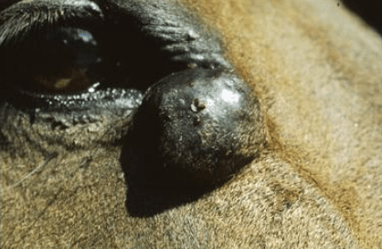
Sarcoids can occur just about anywhere on the body but are most commonly found on the head, (especially around the eyes), the underneath of the abdomen and sheath, chest, ears and lower limbs. Single tumors or a number of sarcoids may occur in one area or over many parts of the body.
6 forms of sarcoids have been described and can vary widely in appearance
1. Occult: appear as round to oval, flat areas of roughened hairless, irregular skin. The skin feels slightly thickened
2. Verrucose: these are ‘wart’ like lesions that commonly occur around head and groin. They will often have areas of hair loss and thickened skin around them.
3. Nodular: have a lumpy appearance.
4. Fibroblastic: are irregularly round, raised, firm lumps. They are usually smooth and hairless at least over part of their surface but smaller ones are sometimes covered with normal-looking skin. If the surface becomes damaged, or often after normal growth, the tumor will ulcerate and bleed, leading to scab formation.
5. Mixed: these sarcoids are common and are composed of a mixture of the above types of sarcoid.
6. Malevolent: rare, highly aggressive form that spread rapidly.
 Visual recognition is the safest and easiest method to diagnose sarcoidosis. The presence of more than one lesion that looks like a sarcoid is strongly supportive of the diagnosis. The only definitive diagnosis is to take a biopsy of the suspected sarcoid and send it to a laboratory; however any biopsy technique carries a high risk of triggering a sudden expansion or change in the nature of the sarcoid. For this reason biopsies of suspected sarcoid lesions are not usually undertaken. Sarcoids can grow to become very large (over 8-10cms, 3.25-4 inches), although most remain smaller than this.
Sarcoids can be similar in appearance to other skin tumors (e.g. fibromas, mast cell tumors and non-pigmented melanomas) and it is necessary to submit a sample (biopsy) or the whole tumor to a laboratory for analysis for a precise diagnosis to be made.
Why do sarcoids occur?
There is strong evidence to support the view that sarcoids are initiated by a virus infection. This theory explains how sarcoids first appear and how they spread. Some genetic families appear particularly susceptible to developing sarcoids more readily than others, but there is no difference in susceptibility between horses of different coat colors. Some breeds may be more susceptible than others. It is not uncommon for ‘proud flesh’ i.e. the exuberant granulation tissue that often develops in healing equine wounds, to transform into sarcoids.
What treatments are available?
There are many suggested treatments, but no single therapy is universally effective. Each case will be assessed individually and the treatment route decided upon will depend on the number of lesions and where these lesions are located amongst other factors. In certain cases, photographs of sarcoids may be sent off for specialist opinion. The important thing to remember is that sarcoids have a great tendency to recur either at the site of removal or nearby. The choice of treatment will depend upon several factors:
- The number and size of the sarcoids present
- The part of the horse affected
- The facilities and drugs available
- Financial considerations
What methods of treatment are there?
- Surgical removal
If is often possible to remove a sarcoid by simply cutting around it after desensitization with local anesthetic and stitching the resulting wound. This is easily done if there is only a solitary tumor or there are only a small number present and there is enough free skin left afterwards to close the wound. Tiny sarcoids may be removed, leaving a small open wound to heal by granulation. Approximately 50% of sarcoids treated this way re-grow subsequently. Surgical removal without additional therapy is not recommended. - Applying ligatures
It is possible to remove the bulk of some sarcoids, especially those with a short stalk or neck, by fixing a tight ligature around its base. The ligature cuts off the tumor’s blood supply and it dies away or falls off usually 10 days to two weeks later. This method is useful for short-term control of relatively large sarcoids on the inside of the hind limbs or abdomen but does not usually give long term resolution of the problem. The most common system used is the application of small strong rubber rings (elastrator rings) using a special applicator. There may be some local swelling after their application but this usually subsides once the sarcoid drops off. Not recommended. - Freezing (Cryosurgery)
The sarcoid may be frozen by using liquid nitrogen or another appropriate freezing agent, which causes the tissue to die away. If the sarcoid is large, most of it can be cut away first (debulking), leaving only the base to be frozen. This method is more effective at preventing recurrence than surgery alone, but often results in the development of patches of white hair due to damage to hair follicles. - Laser surgery
Where available, surgical laser treatment allows the bulk of the sarcoid to be removed and the base eroded either in one step or the base eroded after de-bulking the main mass. There is minimal bleeding because the tissues are burnt, but healing can be slow. Scars will form but hair colour is usually unaffected. - Radioactive beads or wires
This highly specialized technique is not widely used but can be effective particularly for eyelid sarcoids where it is necessary to try to save the eyelid. The radioactive treatment shrinks the tumor and may disfigure the eyelid. - BCG vaccine
BCG is a vaccine produced from the bacterium Mycobacterium bovis for immunization against tuberculosis. It may be injected into the sarcoid tumor(s), often with useful results. Several injections over several weeks or months may be required. This treatment is aimed at provoking an immune reaction from the horse’s body to destroy or reject the sarcoid tissue. It is most commonly used for eyelid tumors because, if effective, it allows the eyelid to be saved. A response may not be seen for several weeks after first injection. There is often initial swelling and there may be skin damage following injections. It is rare but death has been reported following an anaphylactic shock reaction to the vaccine. Horses to be treated with BCG should receive anti-inflammatory drugs prior to each treatment. - Chemotherapy
Specially-prepared cytotoxic (tissue killing) creams have been widely used to treat sarcoid tumors. These attack the abnormal cells in the sarcoid and are often highly effective, but can also damage healthy tissues. They must be used with great care, especially over bony areas or blood vessels and nerves. They can be used on smaller and flat sarcoids or larger ones after surgical debulking. The cream can only be supplied to and used by a veterinarian. Another cytotoxic drug (Cisplatin) is available but must be injected into a sarcoid to be effective. This is another highly specialized technique as dose and pattern of injections varies with size and shape of the sarcoid. Both techniques cause local inflammation and scaring is variable, depending on the size and location of the sarcoids. No matter which treatment option is chosen it can take many months to remove some sarcoids and the effect might not be permanent. Treatment may need to be repeated or changed if new sarcoids appear. Such treatment can be costly.
Should I buy a horse with sarcoids?
Sarcoids affect the potential value of a horse or pony in two main ways:-
1. If they interfere with tack or are knocked during exercise, they reduce the ability of that horse or pony to perform. If a mare has sarcoids between her back legs or on her udder they might be knocked or sucked when the foal nurses.
2. They may be expensive to remove or be treated if prolonged or repeated treatments are required. The above must be considered against the value of the horse and its other qualities or potentials.
Conclusion
Sarcoids are much more significant than ‘just a few lumps’ and can be difficult and costly to deal with. If you think your horse or pony may have one or more sarcoids, you should ask your veterinarian for advice. Best results are achieved when a diagnosis is made and appropriate treatment is started early. Scarring is less obvious when the sarcoids are removed or treated when they are small.